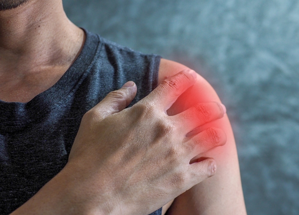
Shoulder pain is one of the most common musculoskeletal complaints in primary care and physiotherapy settings. Rotator Cuff tendinopathy is often the first diagnosis considered, given its prevalence and classic presentation of pain on overhead movements. However, not all shoulder pain follows the expected patterns. Early recognition of alternative diagnoses can be critical in ensuring patients receive the most appropriate care without unnecessary delays.
In this mini-case study, we share an example from our clinic where shoulder pain had an unexpected cause, highlighting the importance of a thorough clinical assessment, targeted imaging, and timely referral.
Patient Presentation
A 42-year-old recreational tennis player presented with a six-week history of gradually worsening right shoulder pain.
The discomfort was primarily located on the lateral aspect of the shoulder and was exacerbated by overhead strokes and lifting objects. There was also some pain in the posterior aspect of the shoulder.
The patient reported stiffness in the morning but denied any trauma, systemic symptoms, or previous shoulder pathology.
On examination:
- Active range of motion was mildly reduced in abduction and forward flexion.
- Pain was reproduced with resisted shoulder abduction and external rotation.
- Palpation over the subacromial space was tender, but there was no obvious swelling or deformity.
- Rotator cuff strength was preserved, and neurological examination was normal.
Based on these features, the initial impression was rotator cuff tendinopathy, a common diagnosis in active adults.
However, subtle atypical features warranted a broader assessment and diagnostic imaging.
Differential Diagnosis Considered
While rotator cuff tendinopathy is common, several other conditions can present similarly. In this case, the differential included:
Glenohumeral Joint Osteoarthritis
Usually presents with deep, diffuse pain and reduced range of motion. Less common in middle-aged active adults without prior trauma.
Labral Tears (SLAP Lesion)
Often associated with clicking, catching, or instability. Common in overhead athletes.
Acromioclavicular Joint Pathology
Pain localized to the AC joint, often exacerbated by cross-body adduction.
Cervical Referral / Nerve Root Irritation
Usually accompanied by radiating pain, numbness, or weakness in a dermatomal pattern.
Calcific Tendinopathy
Severe pain may occur acutely, often with restricted movement; X-ray or ultrasound is diagnostic.
Less Common: Suprascapular Nerve Entrapment / Intra-articular Cysts
Can mimic rotator cuff pain but often presents with subtle atrophy or weakness.
Investigations
Given the atypical features, particularly the absence of significant weakness, the persistence of pain despite conservative measures, and the patient’s high functional demand, we proceeded with imaging:
Xray
No evidence of bony injury found.
MRI
Revealed a small ganglion cyst arising from the spinoglenoid notch, compressing the suprascapular nerve. This was the likely cause of the patient’s shoulder pain and subtle weakness in external rotation.
Diagnosis
Spinoglenoid notch with suprascapular nerve entrapment.
This case highlights the importance of considering nerve-related pathology when standard imaging and clinical findings do not match the expected pattern.
Treatment Approach
The patient’s management and treatment options were discussed in detail.
Conservative Measures
Initial activity modification and physiotherapy focused on gentle strengthening and scapular stabilization.
Non-steroidal anti-inflammatory medication (NSAIDs) was used to control pain.
Specialist Intervention
- Due to ongoing pain and functional limitation, the patient underwent ultrasound-guided aspiration of the ganglion cyst, followed by a targeted physiotherapy programme.
- The procedure was minimally invasive and carried a low risk profile.
Rehabilitation
- Progressive rotator cuff and periscapular strengthening.
- Gradual return to tennis-specific movements over 6–8 weeks.
- Education on load management and early recognition of recurrent symptoms.
Surgical Option
Surgical intervention would likely have been an arthroscopic excision with decompression of the suprascapular nerve. This was discussed, however the patient opted for a less invasive procedure as a first measure.
Outcomes
Within four weeks of the procedure:
- The patient reported significant pain reduction.
- Active range of motion returned to baseline, and overhead activities were pain-free.
- By eight weeks, the patient had resumed full participation in recreational tennis.
Follow-up imaging confirmed resolution of the cyst with no recurrence. This case highlights how early identification of atypical causes can prevent prolonged symptoms and unnecessary interventions such as corticosteroid injections for presumed tendinopathy.
Key Takeaways for Referrers
Not All Shoulder Pain is Tendinopathy
Persistent pain despite appropriate conservative care, or atypical clinical features, should prompt reconsideration of the diagnosis.
Consider Nerve-Related Pathology
Suprascapular nerve entrapment and ganglion cysts can mimic rotator cuff pain. Subtle weakness in external rotation or shoulder atrophy may be clues.
Targeted Imaging Matters
Ultrasound is excellent for assessing tendons and bursae.
MRI is valuable for detecting intra-articular pathology or nerve compression that may not be apparent on initial assessment.
Early Referral Improves Outcomes
Early specialist review can reduce symptom duration, improve function, and avoid unnecessary interventions.
Multidisciplinary Approach is Key
Collaboration between orthopaedic surgeons, radiologists, and physiotherapists helps to ensure accurate diagnosis and a structured rehabilitation pathway.
Conclusion
This mini-case study demonstrates that shoulder pain is not always what it seems. While rotator cuff tendinopathy remains a common diagnosis, clinicians should maintain a high index of suspicion for less frequent conditions such as nerve compression by a ganglion cyst, labral pathology, or other intra-articular lesions.
Timely investigation, specialist input, and a structured rehabilitation plan can lead to excellent functional outcomes and patient satisfaction.
Referrers, whether GPs or physiotherapists, play a crucial role in recognising atypical features early, initiating appropriate imaging, and making relevant referrals to ensure patients receive the care they need.
At London Bridge Orthopaedics, we are committed to supporting referrers with up-to-date clinical guidance, case discussions, and clear referral pathways.
By sharing cases like this, we hope to improve early recognition of complex shoulder presentations and optimise patient outcomes.