What is Hip Impingement?
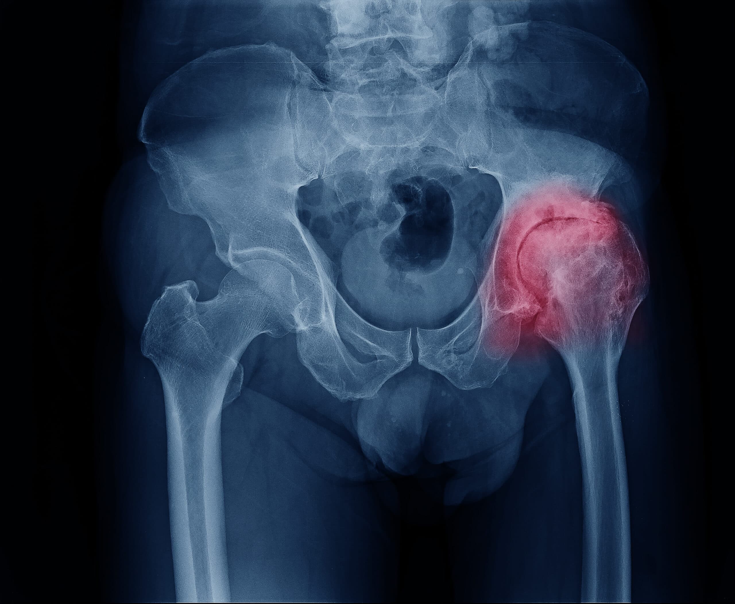
Femoro-acetabular impingement, otherwise called hip impingement, is a relatively newer diagnosis. Because of the newer diagnosis, it has been understood only recently. This has surfaced from the observation that some patients develop early arthritis in their hips because of the abnormality in the shape of the hip bone causing excessive wear. The importance of the recognition of hip impingement lies in the fact that appropriate intervention before the onset of arthritis may stop this from developing later in life, or at least delay the onset.
Hip Joint Components
The hip joint is composed of a ball-and-socket. It is formed by a ball-shaped piece of bone located at the top of your thigh bone called the femoral head. A socket in your pelvis called acetabulum into which the femoral head fits. Attached to the rim of the socket is a washer-like cartilaginous ring called the labrum. The hip is a flexible and stable joint and does not show any abnormal wear stresses during normal range of motion.


What Causes Hip Impingement?
In hip impingement, there is an unwanted contact between the femoral head and acetabulum socket during normal range of hip motion. It is usually due to excessive bony growth at the front part of the femoral head that impinges on the front edge of the socket during normal activities, damaging the labrum and joint cartilage. This type is called cam impingement. The hip impingement can also be due to the presence of a deep socket, called pincer impingement.


This leads to repetitive rubbing injury by the bone prominence. This results in damage to the labrum and the joint lining cartilage and, ultimately, early onset arthritis.
Hip Impingement Symptoms?
Hip impingement commonly presents in younger adults as a deep intermittent groin discomfort of aching pain during or after activity. This is generally noted during or after repetitive or persistent hip flexion or during high-impact physical activities. Sprinting or kicking sports, ascending hills or stairs, prolonged sitting in low chairs, driving or getting in and out of low vehicles exacerbate hip symptoms. There may also be mechanical symptoms of catching or giving way – particularly if there is a treat of the labrum. The hip joint also develops some degree of stiffness, particularly the rotation during hip flexion. During the later phase, the pain becomes constant and is noted even by routine activities.
Diagnosing Hip Impingement
Standard hip X-rays may not always identify the hip impingement condition reliably. A CT or MRI scan of the hip joint is routinely performed for a definitive diagnosis. An MRI arthrogram (MRI scan performed after injecting contrast fluid into the hip joint) is done to assess the associated cartilage damage in detail.
Treatment of Hip Impingement
Hip impingement is a mechanical problem that usually requires a surgical solution. However, targeted sports physiotherapy that focuses on maintaining core stability and a more upright stance might improve the hip symptoms in some patients.
The surgery involves reshaping the hip bones by removing the abnormal bone prominence and repair the damaged cartilage. The aim of the surgery is to improve pain and range of movement in the hip while preventing or stopping the progression of arthritis in the joint. This will depend a lot on the amount of damage that may have already occurred and varies wildly between patients. It is usually possible to address the abnormality through keyhole surgery (hip arthroscopy), but open surgery may be required for specific anatomical abnormalities.
Hip Arthroscopy Surgery
The surgery is generally performed under general anaesthetic. The ball and socket parts of the hip joint are separated by applying traction to the leg with a special device. This provides enough space in the hip joint to allow insertion of a small telescope camera (arthroscope) into the joint safely. Further, one or two small incisions are made to insert special instruments that allow carrying out of the surgical procedure. The procedure is carried out under the guidance of X-ray control.
The duration surgery will vary depending on the type of pathology in the hip joint. This can last from 45 minutes to 120 minutes. At the end of the procedure, a local injection is given around the incisions to minimise pain after the surgery. These keyhole incisions are closed with stitches and covered with a sterile padded dressing.
Recovery After Hip Arthroscopy
The patient usually feels some discomfort in the operated hip area that generally responds well general pain relief medication. There will be some swelling in the groin, buttock and thigh, but that usually resolves within a few days. The patient is mobilised through crutches for 2-4 weeks. Physiotherapy is commenced on the ward soon after surgery and the patient is usually discharged on the same day or next day along with a gentle home exercise regime.
The sports physiotherapist will develop an appropriate outpatient rehabilitation programme according to Mr Kavarthapu’s protocol. The majority of patients walk relatively pain-free 6 weeks after surgery.
Medium impact exercise, such as jogging, can usually be commenced by this time and can gradually progress to running. It may take about 4 to 6 months to return to an elite level of competition/fitness.
The broad strategy for rehabilitation is to regain early range of movement and stability, followed by strength and endurance. Returning to work will depend on pain levels and the nature of your job.
For more information regarding Hip Impingement treatment and all other orthopaedic treatments, contact London Bridge Orthopaedics. For more orthopaedic news, follow us on Facebook, LinkedIn and Twitter.


