Overview of Hip Impingement
Hip impingement syndrome (femoroacetabular impingement or FAI) is caused by unwanted contact between the periphery (outside edge) of the ball and socket parts of the hip joint, which results in damage to the joint cartilage and a decreased range of hip movement.
This hip condition is usually caused by excessive bony growth at the front of the femoral bone (‘bump’) that rubs on the front edge of the socket, damaging the labrum and joint cartilage, even during normal range of motion. This is thought to cause premature arthritis in the hip joint. The patient typically experiences sharp pain during deep hip flexion/bending and twisting movements.
Anatomy of the Hip
The hip joint is one of the largest joints in your body. It is a ball-and-socket joint, formed by the head of the femur (thigh bone) and the acetabulum (socket) of the pelvis. In a normal joint the ball part of the joint fits neatly into the socket in your pelvis.
The bones of the joint are covered by a tough but smooth, slippery surface, known as cartilage. This tissue allows the bones to move against each other without friction.
There are also bands of tissue, called ligaments, that connect the ball to the socket, stabilising the hip and forming the joint capsule. The joint capsule is lined with a thin membrane called synovium, which produces a clear fluid that helps to lubricate the joint.
Fluid-filled sacs called bursae provide extra cushioning where there is friction between muscle, tendons and bones.
The hip is surrounded by large muscles supporting the joint and enabling movement. They include:
- Gluteal muscles – muscles of the buttocks, located on the back of the hip
- Adductor muscles – muscles of the inner thigh, which pull the leg inward toward the opposite leg
- Iliopsoas muscle – a muscle that begins in the lower back and connects to the upper femur
- Quadriceps – four muscles on the front of the thigh that run from the hip to the knee
- Hamstrings – muscles on the back of the thigh, which run from the hip to just below the knee
Major nerves and blood vessels also run through the hip. These include the sciatic nerve at the back of the hip and femoral nerve at the front of the hip, and the femoral artery, which begins in the pelvis and passes by the front of the hip and down the thigh.
This type of joint offers a wide range of motion, provides good stability to carry your body weight, and allows you to move your legs when you are walking, running and jumping.
Causes of Hip Impingement
Hip impingement is often caused by structural abnormalities in the hip joint, leading to friction between the femur and acetabulum. Common causes include:
- Cam Impingement: Excess bone on the femoral head causes friction during hip flexion.
- Pincer Impingement: Overgrowth of the acetabulum rim causes impingement during hip movement.
- Mixed Impingement: A combination of both cam and pincer impingement.
Symptoms of Hip Impingement
The symptoms of hip impingement can vary but often include:
- Hip Pain: Pain in the groin or hip that worsens with activity and is often felt during flexion or rotational movements.
- Limited Range of Motion: Difficulty with hip movements, especially flexion and internal rotation.
- Stiffness: Feeling of stiffness or discomfort in the hip joint.
- Clicking or Snapping: Audible clicking or snapping sensations during hip movement.
Diagnosis of Hip Impingement
Diagnosing hip impingement typically involves:
- Physical Examination: Assessment of hip range of motion, pain, and provocative manoeuvres.
- Imaging: X-rays and MRI scans to visualise the hip joint’s anatomy and detect structural abnormalities.
Treatment Options for Hip Impingement
Treatment for hip impingement aims to relieve pain, improve function, and prevent further joint damage:
- Conservative Management: Rest, activity modification, physiotherapy to strengthen hip muscles, and anti-inflammatory medications.
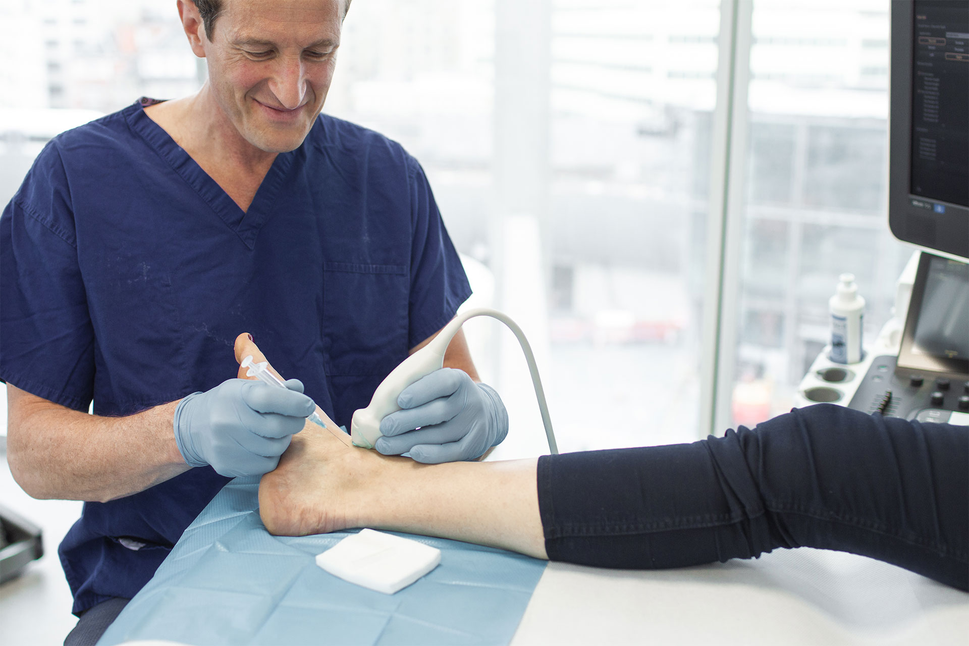
- Injections: Corticosteroid injections can provide temporary pain relief.
Surgical Options
Surgical intervention may be considered, but only if conservative treatments have been ineffective or if there is significant joint damage:
- Hip Arthroscopy: A minimally invasive procedure to remove excess bone and repair damaged tissue in the hip joint.
- Open Hip Surgery: In cases of severe impingement or complex abnormalities, open surgery may be required.
Questions and Answers
Can hip impingement be prevented?
While some factors like anatomy and genetics cannot be changed, maintaining a healthy weight, proper warm-up, and avoiding overuse of the hip joint may help reduce the risk of hip impingement.
How is hip impingement diagnosed?
Diagnosis involves a physical examination, medical history review, and imaging tests such as X-rays and MRI scans.
Can physiotherapy help with hip impingement?
Yes, physiotherapy can improve hip muscle strength, flexibility, and joint mechanics, which may alleviate symptoms and improve function.
What are the potential complications of hip impingement surgery?
Complications can include infection, nerve or blood vessel damage, blood clots, or limited improvement in symptoms. Your surgeon will discuss potential risks before the procedure.
How long does it take to recover from hip impingement surgery?
Recovery time varies based on the surgical approach and individual factors. Patients typically undergo physiotherapy and rehabilitation for several months to achieve full recovery and optimal outcomes.